2D ECHO
2D Echocardiography
The heart is a two-stage electrical pump that circulates blood throughout the body. The anatomy includes four chambers and four valves. For the heart to function normally these structures need to be intact and the heart muscle needs to beat in a coordinated fashion, so that blood flows in and out of each chamber in the proper direction.
The arteries carrying blood to the heart get clogged with time due to the high levels of lipids in the blood and also due to different lifestyle habits such as smoking, drinking, obesity and inactive lifestyle. Once the blockage in the artery reaches 60%-70% the chance of heart attack increases with it. The heart muscle becomes fatty with time and its capacity to pump blood decreases.
Echocardiography is the most common non invasive test for the health of your heart. It displays vital information about the condition of your arteries and the capacity of your heart to pump blood each time it beats.
Uses of Echocardiography
Echocardiography can detect many heart problems. Some may be minor and pose no risk to you while others can be signs of serious heart disease. Echocardiography at Narayan Swaroop Super Speciality Hospital is done to find the following things:
- The size of your heart An enlarged heart might be the result of high blood pressure, leaky heart valves or heart failure. Echo can also detect increased thickness of the ventricles (the heart’s lower chambers).
- Weak Heart muscles That is not pumping blood perfectly. Damage from a heart attack may result in weak areas of heart muscle. Weakening may also mean that the area isn’t getting enough blood supply, a sign of coronary heart disease.
- Heart valve problems Echo can show whether any of your heart valves don’t open normally or close tightly.
- Problems with your heart’s structure Echo can detect congenital heart defect like hole in the heart. Congenital heart defects are structural problems present since the birth. Infants and children may also have an echo test to detect these heart defects.
- Blood clots or tumors If you have had a stroke, you may have to get echo done to check for blood clots or tumors that could have caused the stroke.
Uses of Echocardiography
Other than wearing loose-fitting clothes, there are no special restrictions or preparation prior to a 2D Doppler echocardiogram. During the echocardiogram a gel like material is applied to your chest. Although the gel is water soluble and should not stain; it is messy and could get on your clothing so please be aware and dress appropriately.
Test Results
The purpose of the echocardiogram is to evaluate the structure and function of the heart. The result provides detailed information that can help the health care professional makes a diagnosis related to the heart.
Echocardiograms may be repeated over time, monitoring heart function and the test results may help decide whether previous treatment has been effective or any changes in that treatment program is required.
Heart Checkups
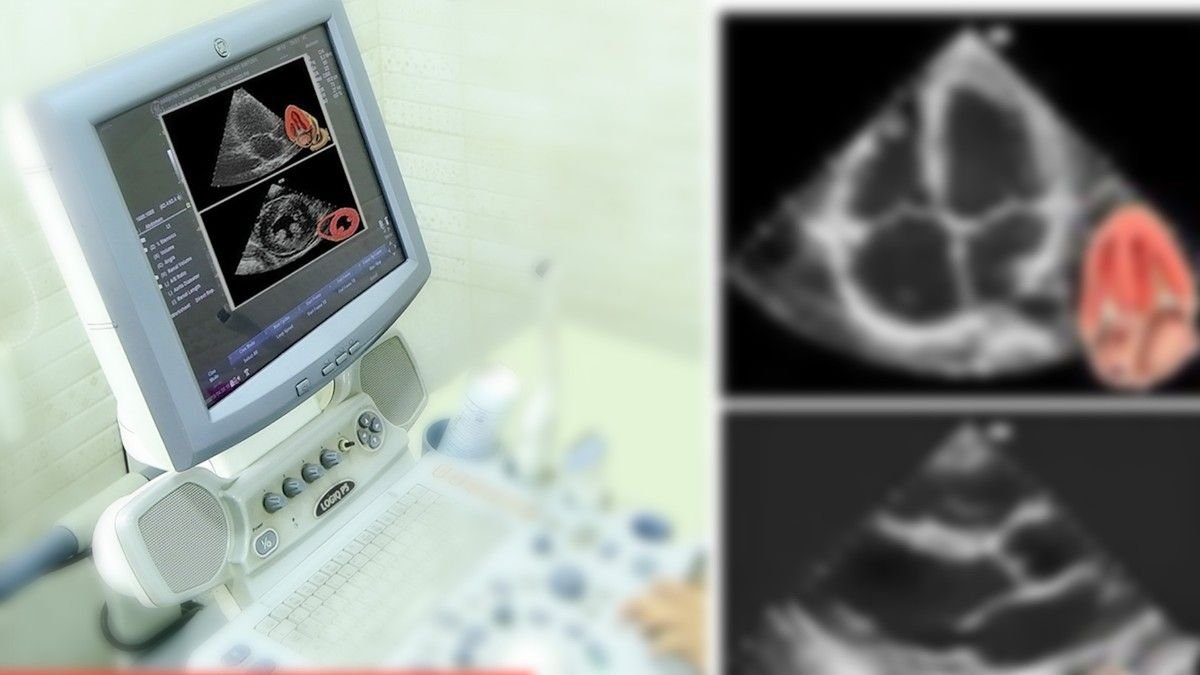
An echocardiogram (echo) is a graphic outline of the heart’s movement. During an echo test, ultrasound (high-frequency sound waves) from a hand-held probe which is placed on your chest provides pictures of the heart’s valves and chambers that help a cardiologist in evaluating the pumping action of the heart. Echo is often combined with Doppler ultrasound and color Doppler to evaluate blood flow across the heart’s valves.
The test is used to:
- Evaluate the overall function of your heart.
- Determine the presence of different types of heart diseases such as valve disease, myocardial disease, pericardial disease & infective endocarditic.
- Cardiac mass & congenital heart disease.
- Follow the progress of valve disease over time.
- Evaluate the effectiveness of your medical or surgical treatments.